
Reflex sympathetic dystrophy (RSD) is a condition that is often described under various synonyms that point to its incompletely understood etiology. In 1864, Weir Mitchell coined the term causalgia to designate severe pain following nerve injury. In 1900, Sudeck described regional demineralization accompanying posttraumatic pain. In 1923, Leriche described vasomotor disequilibrium.[1] In 1947, Evans introduced the term reflex sympathetic dystrophy.
In 1993, the International Association for the Study of Pain renamed algodystrophy complex regional pain syndrome (CRPS; also known as chronic regional pain syndrome). RSD is type 1 CRPS. It can be considered an excessive sympathetic reaction of joints and periarticular soft tissues to any insult, traumatic or unknown.[2] This is quite different from causalgia (type 2 CRPS), in which the etiology is a partial nerve injury.
RSD is characterized by pain, regional edema, joint stiffness, muscular atrophy, vasomotor disturbances (including temperature changes), trophic skin changes, and regional skeletal demineralization seen on radiographs. These changes are aggravated by activity and extend over a larger area than the primary injury or surgery, including the area distal to this focus.[3, 4, 5, 6]
Because pathognomonic criteria are lacking for RSD, a taxonomic system based on clear definitions and objective quantification is desirable. Therefore, the current terminology of CRPS is increasingly being used as an umbrella to replace the myriad empirical descriptions used previously. No apparent relation exists between the degree of initial trauma and the severity of RSD, but RSD generally is more frequent following minor trauma or operations.
NextRSD usually follows minor trauma or surgery. It also has been associated with various clinical conditions (eg, diabetes, parkinsonism). It begins with spontaneous pain associated with vasomotor and sudomotor disturbances.
Bonica described the progress of severe cases of RSD in three stages, as follows[7] :
Iatrogenic causes of RSD following surgery, such as carpal tunnel decompression or Dupuytren release, can be diagnosed easily.[8] No clear etiology (including trauma) can be identified in 25-35% of cases. A detailed history can be useful to pinpoint uncommon causes of RSD.
Potential causes of RSD include the following:
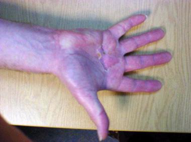
 Reflex sympathetic dystrophy following surgery for Dupuytren contracture.
Possible contributory causes include the following:
Statistically significant associations include the following:
According to various researchers, the incidence of RSD may be 2-17% following minor trauma or surgery. If causalgia is included in the broad definition, incidence can be as high as 32-35%. In the past, many subtle forms of RSD were missed, but with increased awareness of the condition, actual incidence may be much higher than initially thought.
Worldwide, no regions or population groups have been demonstrated to have a predilection for RSD.
Most patients with RSD are aged 30-55 years; the mean age is 45 years. With increasing awareness, RSD is being diagnosed in children more often; however, no studies exist pointing to a particular age distribution.
RSD is more common in women than in men; the male-to-female ratio is approximately 3:7. The ratio of upper-extremity to lower-extremity involvement is approximately 2:1. Even in children, girls are affected more frequently than boys, but peculiarly, the lower extremity is involved more frequently than the upper extremity.[13]
No particular race has a predilection for reflex sympathetic dystrophy.
Mortality associated with RSD is negligible, though morbidity is extremely high. About 80% of patients with RSD diagnosed within 1 year of injury improve significantly. However, 50% of patients with untreated symptoms lasting more than 1 year have profound residual impairment.
Despite good results following intravenous sympathetic blockade and intensive mobilization techniques, weakness of the extremity resulting from RSD is seen in almost 50-65% of patients, even 18-24 months after the initial diagnosis. Full range of movement accompanying the above aggressive therapies is seen in 60-74% of patients. Prolonged morbidity is observed in about 50% of patients with psychiatric diathesis, workers' compensation claims, and lawsuits.[14]
Emphasize to all patients the importance of early and supervised mobilization of the affected part. Give patients a list of plaster instructions and a list of realistic goals to be achieved within a specified time interval. Any lag in achieving these objectives due to pain, swelling, or other causes should inspire concern leading to early diagnosis and institution of treatment.
Clinical Presentation
Copyright © www.orthopaedics.win Bone Health All Rights Reserved