
Spondylolisthesis refers to the forward slippage of one vertebral body with respect to the one beneath it. This most commonly occurs at the lumbosacral junction with L5 slipping over S1, but it can occur at higher levels as well. It is classified on the basis of etiology into the following five types[1] :
Many cases can be managed conservatively. However, in persons with incapacitating symptoms, radiculopathy, neurogenic claudication, postural or gait abnormality resistant to nonoperative measures, and significant slip progression, surgery is indicated. The goal of surgery is to stabilize the spinal segment and decompress the neural elements if necessary.[2, 3, 4, 5, 6, 7, 8] (See the images below.)
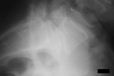
 Spondylolisthesis, spondylolysis, and spondylosis. Isthmic spondylolisthesis (type IIa) with grade 2 slippage of L5 over S1 and spondylolysis (lytic pars defect) is depicted posteriorly.
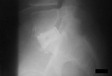
 Spondylolisthesis, spondylolysis, and spondylosis. Although interbody devices afford immediate stability to the anterior column, their use as stand-alone devices has been associated with pseudoarthrosis. Thus, concomitant posterior fixation is often used to augment their stability.
For more information on this topic, see Spondylolisthesis Imaging, Spondylolysis Imaging, Lumbar Spondylosis, Diagnosis and Management of Cervical Spondylosis, and Lumbosacral Spondylolysis.
NextIn 1854, Killian coined the term spondylolisthesis to describe the gradual slippage of the L5 vertebra due to gravity and posture. In 1858, Lambi demonstrated the neural arch defect (absence or elongation of pars interarticularis) in isthmic spondylolisthesis. Albee and Hibbs separately published their initial work on spinal fusion. Their methods were applied quickly to cases involving trauma, tumors, and, later, scoliosis. In the latter half of the 20th century, spinal fusion was used increasingly to treat degenerative disorders of the spine, including degenerative spondylolisthesis and degenerative scoliosis.
Spondylolisthesis is the forward slippage of one vertebra on another. This may or may not be associated with gross instability of the spine. Some individuals remain asymptomatic even with high-grade slips, but most complain of some discomfort. It may cause any degree of symptoms, from minimal symptoms of occasional low back pain to incapacitating mechanical pain, radiculopathy from nerve root compression, and neurogenic claudication.
The incidence of isthmic type (see Etiology for definition) of spondylolisthesis is believed to be approximately 5% based on autopsy studies.
Degenerative spondylolisthesis is observed more frequently as the population ages and occurs most frequently at the L4-L5 level. Up to 5.8% of men and 9.1% of women are believed to have this type of listhesis.
The etiology of spondylolisthesis is multifactorial. A congenital predisposition exists in types 1 and 2, and posture, gravity, rotational forces, and high concentration of stress loading all play parts in the development of the slip.
The following scheme of spondylolisthesis types, based on etiology, is adapted from Wiltse et al[1] :
Spondylolisthesis can be graded based on the amount of vertebral subluxation in the sagittal plane, as adapted from Meyerding (1932):
The dysplastic type occurs from a neural arch defect in the upper sacrum or L5. In this type, 94% of cases are associated with spina bifida occulta. A high rate of nerve root compression at the S1 foramen exists, although the slip may be minimal (ie, grade 1).
The pars interarticularis, or isthmus, is the bone between the lamina, pedicle, articular facets, and the transverse process. This portion of the vertebra can resist significant forces during normal motion. The pars may be congenitally defective (eg, in spondylolytic subtype of isthmic spondylolisthesis) or undergo repeated stress under hyperextension and rotation, resulting in microfractures. If a fibrous nonunion forms from ongoing insult, elongation of the pars and progressive listhesis results. This occurs in the second and third subtypes of type 2 (isthmic) spondylolisthesis. These typically present in the teenage or early adulthood years and are most common at L5-S1.
A unilateral pars defect (spondylolysis) may not demonstrate any degree of slippage; thus, a patient may have spondylolysis without spondylolisthesis. The reverse is also true as in the degenerative-type slips described below.
Biomechanical factors are significant in the development of spondylolysis leading to spondylolisthesis. Gravitational and postural forces cause the greatest stress at the pars interarticularis. Both lumbar lordosis and rotational forces are also believed to play a role in the development of lytic pars defects and the fatigue of the pars in the young. An association exists between high levels of activity during childhood and the development of pars defects. Genetic factors also play a role.
In degenerative spondylolisthesis, intersegmental instability is present as a result of degenerative disc disease and facet arthropathy. These processes are collectively known as spondylosis (ie, acquired age-related degeneration). The slip occurs from progressive spondylosis within this 3-joint motion complex. This typically occurs at L4-5, and elderly females are most commonly affected. The L5 nerve root is usually compressed from lateral recess stenosis as a result of facet and/or ligamentous hypertrophy.
In 2014, the French Society for Spine Surgery proposed a new classification of degenerative spondylolisthesis, comprising the following five types[9] :
In traumatic spondylolisthesis, any part (usually not pars) of the neural arch can be fractured, leading to the unstable vertebral subluxation.
Pathologic spondylolisthesis results from generalized bone disease, which causes abnormal mineralization, remodeling, and attenuation of the posterior elements leading to the slip.
The clinical presentation differs, depending on the type of slip and the age of the patient.
During the early years of life, the presentation is one of mild low back pain that occasionally radiates into the buttocks and posterior thighs, especially during high levels of activity. The symptoms rarely correlate with the degree of slip, although they are attributable to segmental instability. Neurologic signs often correlate with the degree of slippage and involve motor, sensory, and reflex changes corresponding to nerve root impingement (usually S1). Progression of listhesis in these young adults usually occurs in the setting of bilateral pars defects and can be associated with the following physical findings:
The patient with degenerative spondylolisthesis is typically older and presents with back pain, radiculopathy, neurogenic claudication, or a combination of these symptoms. The slip is most common at L4-5 and less common at L3-4. The radicular symptoms often result from lateral recess stenosis from facet and ligamentous hypertrophy and/or disc herniation. The L5 nerve root is affected most commonly and causes weakness of the extensor hallucis longus. Concomitant central stenosis and neurogenic claudication may or may not exist.
The cause of claudication symptoms during ambulation is multifactorial. The pain is relieved when the patient flexes the spine by sitting or by leaning on shopping carts. Flexion increases canal size by stretching the protruding ligamentum flavum, reduction of the overriding laminae and facets, and enlargement of the foramina. This relieves the pressure on the exiting nerve roots and, thus, decreases the pain.
The goal of surgery is to stabilize the segment with listhesis and decompress any of the neural elements under pressure. Restoration of normal sagittal alignment must also be achieved. When evaluating a patient, many factors, such as age, degree of slip, and risk of slip progression, must be considered. Thus, each patient's treatment algorithm should be individualized to achieve optimal outcome.
The indications for spinal fusion clearly differ in the pediatric and adult populations. For the younger population, the following factors are known to correlate with higher risk of slip progression:
However, many young patients are treated with immobilization or activity modification alone, with a significant success rate. In the absence of high-grade slips, minimal symptomatology, and lack of slip progression, fusion is generally not indicated in this population.
Before surgery is considered for adult patients presenting with degenerative spondylolisthesis, minimal neurologic signs, or mechanical back pain alone, conservative measures should be exhausted, and a thorough evaluation of social and psychological factors should be undertaken.
Indications for surgical intervention (fusion) are as follows:
In persons with congenital-type spondylolisthesis, dysplastic articular facets predispose the spinal segment to listhesis due to their inability to resist anterior shear stress. The pars may be intact, or it may undergo microfractures. Thus, it may not be the initiator of listhesis in dysplastic types. The risk of slip progression is high.
The pars interarticularis, or isthmus, resists significant forces during normal motion. The pars may be congenitally defective (isthmic spondylolisthesis is spondylolysis) or undergo repeated stress under hyperflexion and rotation resulting in microfractures. Lumbar lordosis, gravity, posture, high-intensity activities (eg, gymnastics), and genetic factors all play a role in the slip development. If a fibrous nonunion forms from ongoing insult, elongation of the pars and progressive listhesis results; this is observed in another subtype of type 2 (isthmic) spondylolisthesis. In persons with spondylolysis, 30-50% are believed to progress to spondylolisthesis. The most common location is at L5-S1.
Degenerative spondylolisthesis results from intersegmental instability. The pathophysiology of disc degeneration and facet arthropathy has been investigated extensively; however, the nature and etiology of pain generation in the absence of canal or lateral recess stenosis is still debated. Degeneration of the annulus fibrosis results in radial tears through which a posteriorly migrated nucleus pulposus can herniate. Degeneration of the disc may also lead to changes affecting the stability of the spinal motion segment, thus affecting the articular facets. Disc desiccation places greater stress on the facets, which are then subjected to shear forces. The subluxation occurs as a result of progressive facet incompetence. This type most commonly occurs at L4-5 and L3-4.
Surgery is contraindicated if the patient is in poor medical health and if the operative risk is not outweighed by the potential benefits.
Anticoagulation with warfarin, or antiplatelet therapy, can make the risk of hemorrhage much higher than routinely expected. Antiplatelet therapy should be discontinued 3-5 days prior to the procedure. Coumadin should be stopped 5-7 days prior to the procedure, and a prothrombin time result within reference range should be achieved before surgery.
Smoking significantly decreases the chance for a successful fusion. Some surgeons prefer that a patient commit to smoking cessation up to one month before the surgical procedure.
Correction of the listhesis is associated with risk of neurologic injury, both transient and permanent. Some surgeons prefer to fuse the spine in place rather than to reduce the subluxation. In persons with higher-grade spondylolisthesis, use of interbody grafts is associated with a high rate of complications. However, the use of these devices adds to the stability of the spinal segment, helps with the reduction of the deformity, and helps achieve sagittal balance, thus ensuring better outcome.
Workup
Copyright © www.orthopaedics.win Bone Health All Rights Reserved