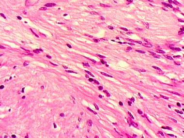

Neurilemmomas (neurilemomas) are benign, encapsulated tumors of the nerve sheath. Their cells of origin are thought to be Schwann cells derived from the neural crest (see the image below). These masses usually arise from the side of a nerve, are well encapsulated, and have a unique histologic pattern.
 The cell of origin for a neurilemmoma is the Schwann cell, which is derived from the neural crest. These cells line the peripheral nerve processes.
This benign lesion essentially manifests itself with cosmetic deformity, a palpable mass, symptoms similar to a compressive neuropathy, or some combination of these. Neurologic symptoms tend to present late. Symptoms can be vague, and there is an average interval of up to 5 years before the diagnosis is established.
NextThe cause of these neoplasms is unknown.[1] Neurilemmoma can be associated with von Recklinghausen disease; when this is the case, multiple tumors often are present.
Neurilemmoma is the most common neurogenic tumor, but precise prevalence figures are not available. They affect persons aged 20-50 years. No racial or sex predilection is recognized. Common locations for the tumors are, in order of decreasing frequency, the head and flexor surfaces of the upper and lower extremities and the trunk.
Recurrence is unlikely after complete resection. Patients usually have rapid and complete relief of pain, with excellent long-term results.[2]
Rare descriptions exist of malignant change in long-standing neurilemmomas, usually in patients with an underlying diagnosis of neurofibromatosis. Malignant change is extremely rare in isolated lesions.
Kano et al evaluated tumor control and hearing preservation relating to tumor volume, imaging characteristics, and nerve and cochlear radiation dose after stereotactic radiosurgery with a Gamma Knife (Elekta, Stockholm, Sweden) in patients with acoustic neuroma.[3]
At a median of 20 months after surgery, none of the patients required further treatment.[3] Serviceable hearing was preserved in 71% of all patients and in 89% of patients with Gardner-Robertson (GR) class I hearing. Patients who received a radiation dose lower than 4.2 Gy to the central cochlea had significantly better hearing preservation of the same GR class, and all 12 patients younger than 60 years who received a cochlear radiation dose lower than 4.2 Gy retained serviceable hearing at 2 years after surgery.
Clinical Presentation
Copyright © www.orthopaedics.win Bone Health All Rights Reserved